View Options
Use these options to customize the view for a reading step in a sequence protocol when using the Protocol Capture tool. Some of the following view options may appear for specific modalities only.
Empty ViewportEnable to force the viewport to be empty regardless of whether the Fill Unmatched Viewports with Non-Displayed Series modality user preference is enabled.
Secondary ImageEnable to display a secondary image. This option works with the matching criteria for Secondary Images. For example, enable this option to display a scanned document. Enabling this option replaces most options with Secondary Images option in the Matching Criteria options.
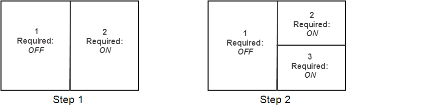
RequiredEnable to show the reading step if the required view or one of the required views is matched. If the step includes more than one required view, the step appears if one of the required views are matched. InteleViewer displays unmatched views as part of the Other Images reading step in the Protocol Preview pane.
For example, for step 1, if a series is not matched with view 2, step 1 will not appear in the sequence protocol. Step 2 appears if a series matches either view 2 or 3, and does not appear if a series match is not found for views 2 and 3.
Matching Criteria
ChronologySpecifies whether the series appearing in the viewport should come from the current or prior study. Choose one of the following options:
|
Option |
Description |
|---|---|
|
Current |
The current study |
|
nth Prior |
A recent prior, arranged from most recent to last. |
|
Oldest Prior |
The oldest prior |
Ultrasound TypeSpecifies the ultrasound (US) view according to the view position. You can choose Scalar, Cine, or Any. Use Any to match any US view. This option is not available when Secondary Image is enabled.
View OrientationSpecifies the view according to the view position. This option is not available when Secondary Image is enabled.
For X-ray, choose one of the following view positions:
|
View Position |
Description |
|---|---|
|
Any |
Match any X-ray view |
|
PA or AP |
Posterior/Anterior or Anterior/Posterior images |
|
Lat |
All lateral images (with a right-lateral and left-lateral orientation) |
|
Decub |
All decubitus images (Right Lateral Decubitus and Left Lateral Decubitus) |
|
Obl |
All oblique images (Right Lateral Oblique and Left Lateral Oblique) |
InteleViewer determines the view position of the X-ray image by matching the DICOM value for the View Position attribute contained in the image to the predefined DICOM values (AP, PA, LL, RL, RLD, LLD, RLO, and LLO). If the image does not have the DICOM View Position data, then InteleViewer searches for these values in the series description: “lat,” “dec,”, or “obl.’ If there is no match, then InteleViewer will not apply the reading step containing that view (excluding Any view).
For CT or MR, choose one of the following view positions:
|
View Position |
Description |
|---|---|
|
Any |
Match any view |
|
Ax |
All Axial images |
|
Sag |
All Sagittal images |
|
Cor |
All Coronal images |
Slice ThicknessSpecifies the slice thickness. You can choose Thinnest, Thickest, or Any. Use Any to match any thickness. This option is not available when Secondary Image is enabled.
InteleViewer determines the slice thickness by matching the DICOM value for the Slice Thickness attribute to an image in the series. If the series has more than one image, InteleViewer uses an image in the middle of the series.
Contrast PresenceSpecifies whether or not a contrast agent is present. You can choose +C (with contrast), -C (without contrast), or Any. Use Any to match any or a particular CT view regardless of whether a contrast agent is present. InteleViewer determines the presence of a contrast agent by matching the DICOM value for Contrast Bolus Agent attribute to that of the images in the series. This option is not available when Secondary Image is enabled.
For example, you open a CT study that contains the following DICOM value for Contrast Bolus Agent:
(0018,0010) ContrastBolusAgent LO #10 1 [OMNIPAQUE]
InteleViewer uses this value as the matching criteria. Since the Contrast Bolus Agent value is equal to “Omnipaque,” InteleViewer determines that this study has a contrast agent. If a sequence protocol contains a reading step that specifies a view with contrast, then only CT series with contrast will match that view. If you open a CT study that contains the DICOM value for Contrast Bolus Agent of “no,” “none,” or “off,” or is empty, then InteleViewer will not apply the sequence protocol to the CT study.
Window LevelInteleViewer determines the window level. This option is not available when Secondary Image is enabled.
The window level is determined by matching the DICOM value for Window Width and Window Level to the values defined in the following table:
|
Window Level |
Matching Criteria |
|---|---|
|
Any |
InteleViewer matches any or a particular CT view regardless of whether the DICOM values for Window Center and Windows Width are specified in the image. |
|
Lung |
If the DICOM value for Window Center is less than or equal to -300, then InteleViewer identifies the study as a lung study. If the DICOM value for Window Center is less than or equal to 0 and the value for Window Width is greater than or equal to 1000, then InteleViewer identifies the study as a lung study. |
|
Bone |
If the DICOM value for Window Center is greater than or equal to 200, then InteleViewer identifies the study as a bone study. If the DICOM value for Window Center is greater than or equal to 100, and the value for Window Width is greater than or equal to 1000, then InteleViewer identifies the study as a bone study. |
|
Soft tissue |
InteleViewer identifies the series as soft tissue in the following scenarios:
|
For example, you open a CT study that contains the following DICOM values for Window Center and Window Width:
(0028,1050) WindowCenter DS #4 1 [80.0]
(0028,1051) WindowWidth DS #6 1 [186.0]
InteleViewer uses these values as the matching criteria. Since the Window Center attribute value is equal to “80,” InteleViewer determines that this study has a window level value of “Soft Tissue.” If a sequence protocol contains a reading step with a view where the Reconstruction Algorithm is set to Soft, then only CT series with a window level value of “Soft Tissue” will match that view.
Use Series DescriptionEnable to create rules. The rule you create is based on the text you enter and the matching operator you specify. The rule will be used to identify the series (such as, localizer or scout images or modality screenshot images) or document. For example, if the series description in the view options specifies the Equals operator and the text “scan req,” and Any is chosen for all other matching criteria, then InteleViewer will match the view to a study containing the series description “scan req.”
You can create multiple rules by adding new lines and selecting additional operators. No more than 50 rules can be combined at once to identify matching series descriptions. Within the maximum of 50, you cannot use more than one Equals operator or more than one Begins With operator.
Setting multiple rules for series description cannot be done using the Protocol Manager, but only by accessing Matching Criteria dialog via View Options.
The series description text is case-insensitive. Blank entries are unusable as match criteria; the rule will be discarded after selecting Done.
If you set matching criteria other than Any, the view matches specific series only. In this case, all non-diagnostic images (generic datasets), such as a scout images and scanned documents, are ignored and will not match the view, regardless of the series description setting.
Choose an operator in the following table to determine how the series description should match the text.
|
Choose: |
To match series descriptions that: |
|---|---|
|
Contains All Of |
Contain all of the specified text, in any order. Use quotation marks to specify exact phrases. |
|
Contains Any Of |
Contain at least one of the specified text, in any location. Use quotation marks to specify exact phrases. |
|
Equals |
Contain all the text exactly as it appears, including spaces and without any additional characters. |
|
Begins With |
Begin with the specified text. Studies with the text located elsewhere in the description (such as in the middle) will not be matched. |
|
Contains None Of |
Do not contain any of the specified text. |
When using the Contains All Of, Contains Any Of, and Contains None Of operators, partial matches are supported—the specified text does not require preceding and following spaces in the study description to match. For example, if the study description contains “abdomen pelvic,” the text “abdo” will match.
SequenceChoose one of the following sequences: T1 (T1-weighted), T1 GRE (T1 Gradient Echo), T2 (T2-weighted), T2 GRE (T2 Gradient Echo), PD (Proton Density), DWI (Diffusion Weighted Imaging), ADC (Apparent Diffusion Coefficient), Exp ADC (Exponential ADC), STIR (Short T1 Inversion Recovery), FLAIR (Fluid Attenuated Inversion Recovery), and Any. Use Any to match any or a particular MR view regardless of whether a sequence is specified in the series description. This option is not available when Secondary Image is enabled.
If you cannot find the required sequence in the MR view option, define a new sequence by using series matching.
InteleViewer determines the sequence by matching the series description to a series in the MR study, as well as matching the DICOM values for flip angle and inversion time to the values defined in the following table:
|
Sequence |
Match Criteria |
|---|---|
|
T1 |
Series description contains T1 as any part of the string (including T1W or T1_Axial) and the flip angle is ≥ 90 degrees. |
|
T1 GRE |
Series description contains T1, FLASH, SPGR, FSPGR, MPSPGR, or FMPSPGR as any part of the string (including T1 GRE, T1 FFE, and T1 CE-FFE) and the flip angle is < 90 degrees. |
|
T2 |
Series description contains T2 and the flip angle is ≥ 90 degrees. |
|
T2 GRE |
Series description contains T2, GRE, FFE, FISP, MPGR, GRASS, FMPGR, or MERGE (including T2 with any additional characters) and the flip angle is < 90 degrees. |
|
PD |
Series description contains PD. |
|
STIR |
Series description contains STIR or the inversion time value is < 250 ms. |
|
FLAIR |
Series description contains FLAIR or the inversion time value is ≥ 250 ms. |
|
DWI |
Series description contains DWI, DW, diffusion, or diff. |
|
ADC |
Series description contains ADC or apparent. |
|
Exp ADC |
Series description contains exponential. |
For example, you open an MR study that contains the series description “SAG T1 FSE-LT” and the following DICOM value for FlipAngle:
(0018,1314) FlipAngle DS # 4 1 [94.0]
InteleViewer uses these values as the matching criteria. Since the FlipAngle attribute value is equal to “94” and the series description contains “T1,” InteleViewer determines that this study has a T1 sequence. If a sequence protocol contains a reading step with a view where the sequence value is equal to “T1,” then only MR series with a FlipAngle attribute value greater than or equal to “90” will match that view.
If there are several sequences that match a series description (for example, SAG T1 FLAIR), InteleViewer will rank the sequences in the following order: T1, T2, PD, T1_GRE, T2_GRE, STIR, FLAIR, DWI, ADC, EXP ADC.
Fat SaturationSpecifies whether or not to match a series based on fat saturation. You can choose No Fat Sat or Fat Sat. This option is not available when Secondary Image is enabled.
If you choose Fat Sat, a series matches if the series description contains FAT SAT, CHEM SAT, SPIR, SPAIR, WaterSEL, FS, FATSAT, FAT_SAT, F/S, or F_S, and does not contain NO FATSAT, NO FS, NOFS, WOFS, NO_FS, WO_FS, WO/FS, or NO/FS.
If you choose No Fat Sat, a series matches if the series description does not contain FAT SAT, CHEM SAT, SPIR, SPAIR, WaterSEL, FS, FATSAT, FAT_SAT, F/S, or F_S.
If you open an MR study that contains the text “FSE” or “FSPGR,” InteleViewer will not apply the sequence protocol to the MR study.
Contrast PresenceSpecifies whether or not a contrast agent is present. You can choose +C (with contrast), -C (without contrast), or Any. Use Any to match any or a particular MR view regardless of whether a contrast agent is present. This option is not available when Secondary Image is enabled.
InteleViewer determines the presence of a contrast agent by matching the DICOM value for Contrast Bolus Agent to that of the images in the series.
For example, you open an MR study that contains the following DICOM value for Contrast Bolus Agent:
(0018,0010) ContrastBolusAgent LO #10 1 [OMNIPAQUE]
InteleViewer uses this value as the matching criteria. Since the Contrast Bolus Agent value is equal to “Omnipaque,” InteleViewer determines that this study has a contrast agent. If a sequence protocol contains a reading step with a view where the +C (with contrast) option is selected, then only MR series with a Contrast Bolus Agent value will match that view. If you open an MR study that contains the DICOM value for Contrast Bolus Agent of “no,” “none,” or “off,” or is empty, then InteleViewer will not apply the sequence protocol to the MR study.
3DSpecifies whether or not the series description contains 3D. You can choose No 3D or 3D.
Reformat Options
Enable ReformatsEnable to activate and set the reformat options.
Your reformat settings are maintained when this option is disabled.
PlaneSpecifies the orientation for the view. You can choose Ax (Axial), Cor (Coronal), or Sag (Sagittal). Choose Don't Modify to preserve the original view orientation.
ThicknessSpecifies the thickness for the reformat. Choose Custom, and enter a thickness value from 0 to 50 (in millimeters). Choose Don't Modify to preserve the original thickness.
Slabbing AlgorithmSpecifies the intensity projection of the slice. Choose one of the following algorithms:
|
Algorithm |
Description |
|---|---|
|
AVG (average intensity projection) |
Display the average pixel value for the slab. |
|
MIP (maximum intensity projection) |
Display the maximum pixel value for the slab. |
|
MINIP (minimum intensity projection) |
Display the minimum pixel value for the slab. |
Display Options
Enable Display OptionsEnable to activate and set the display options.
Your display settings are maintained when this option is disabled.
Initial ImageSpecifies the image in the series that is displayed by default. You can choose Beginning of Series, Middle of Series, or End of Series.
Color Map ModeSpecifies the initial grayscale or color map mode of the image. You can choose From DICOM Data, Inverse of DICOM Data, Low Pixel Values as White, or Low Pixel Values as Black.
SortingSpecifies how you want to sort the images in multi-image series. You can choose Sort By Image Number, Sort By Image Position, Sort By Image Temporal Position, or Sort By Image Time.
Viewport ScaleSpecifies the size of the series, as a percentage, relative to the viewport.
Rotation AngleSpecifies the rotation angle of the series in the viewport. You can choose -90°, 0°, 90°, or 180°.
FlipSpecifies whether to flip the series horizontally or vertically. You can choose Horizontal, Vertical, or both to flip the series both horizontally and vertically.
Window LevelAutomatically applies the window level setting to the series in the study. You can choose the window level presets: Lung, Bone, and Soft Tissue, or choose the window level setting from the DICOM data contained in the image. You can also set custom width and level values by choosing Custom.
The Window Level setting is active regardless of if Enable Display Options is enabled or disabled.
By default, InteleViewer applies the following window level settings to CT studies:
|
Preset Name |
Width |
Level |
|---|---|---|
|
Lung |
1500 |
-600 |
|
Bone |
2500 |
480 |
|
Soft Tissue |
350 |
40 |
If the CT modality user preferences include Lung, Bone, or Soft Tissue window level presets, InteleViewer will use these presets as the matching criteria instead of the same presets defined by the view. Furthermore, window level presets in a presentation state will override presets defined by a view.