Measurement Calibration for Projection Radiographic Images
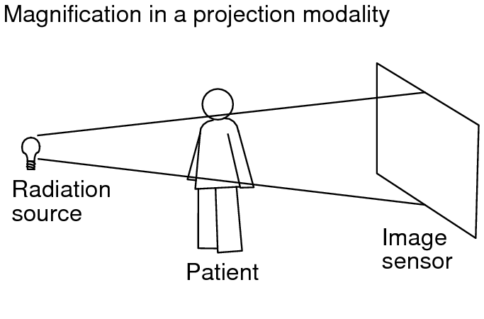
Projection radiographic images that are captured at the scanner are magnified because of the distances between the source of radiation, the patient, and the scanner’s image sensor. The greater these distances, the greater the magnification factor. The diagram below illustrates this magnification effect.
Measurements on a projection radiographic image must be calibrated for the magnification factor in order to display measurements based on the patient's true physical size. In InteleViewer, calibrated measurements are displayed in anatomical units as opposed to measurements based on the imager plate. InteleViewer computes calibrated measurements from a combination of the following three DICOM attributes:
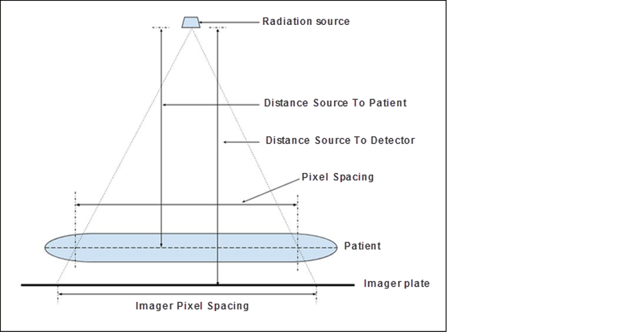
- Imager Pixel Spacing (IPS)The actual distance between pixel centers on the imager plate. The DICOM values are 0018 and 1164.
- Pixel Spacing (PS)The physical distance in the patient between the centers of each pixel. The DICOM values are 0028 and 0030.
- Estimated Radiographic Magnification Factor (ERMF)The calibration value to apply to the Imager Pixel Spacing value so that it is converted to anatomical units. The DICOM values are 0018 and 1114. ERMF is the ratio between the Distance Source To Patient and the Distance Source To Detector.
The IPS value is considered to be the most reliable value for calibrating measurements. When the projection radiographic image includes both the IPS and the PS DICOM attributes, where the IPS value is greater than the PS value, the IPS value is considered to be valid. When the IPS value is less than the PS value, the IPS value is considered to be invalid.
If the DICOM data includes a valid IPS value and an ERMF value, then InteleViewer uses these two values to automatically compute the approximate true physical size of measurements, and displays the measurements in anatomical units.
Depending on the available DICOM data, InteleViewer may use the PS value instead of the IPS value to calibrate measurements. For example, when the image includes a valid IPS value but the ERMF value is absent, InteleViewer uses the PS value to calibrate the measurements.
If there is insufficient DICOM data to compute the true physical size for the measurements, InteleViewer does not calibrate the measurements. For example, when an image does not contain IPS and PS values. For uncalibrated measurements, you can use the Calibrate Measurement tool to manually calibrate the measurements. In this case, InteleViewer uses your calibration to compute the patient's true physical size.
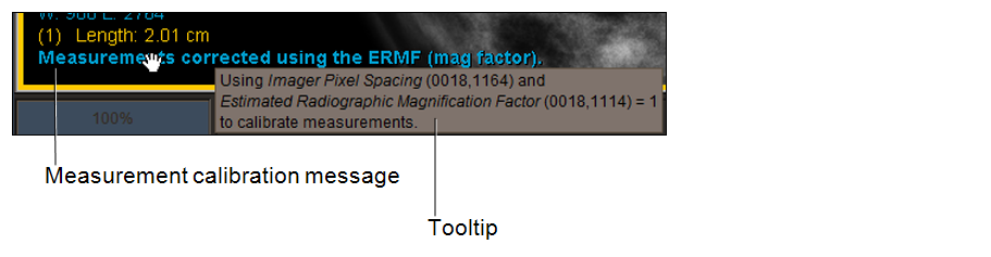
When you take a measurement, a message appears at the end of the list of measurements in the viewport to help you to determine the relevant accuracy of your measurements. You can hover your mouse cursor over the message to display a tooltip that describes the DICOM attributes that are used by InteleViewer to determine measurement calibration.
The following table lists the measurement calibration messages that appear in the viewport, and their corresponding DICOM attribute conditions.
|
Message |
IPS |
PS |
ERMF |
|---|---|---|---|
|
Measurements not calibrated. |
Absent |
Absent |
Ignored |
|
Measurements corrected at modality. |
Valid |
Present |
Absent |
|
Measurements not corrected. Measured size is at detector. |
Valid |
Absent |
Absent |
|
Measurements corrected using the ERMF (mag factor). |
Valid |
Ignored |
Present |
|
Measurements are uncertain. |
Invalid or Absent |
Present |
Absent |
|
Measurements are uncertain. |
Absent |
Present |
Present |
|
Measurements are uncertain. |
Invalid |
Present |
Present |
|
Measurements are user calibrated. This message appears when you use the Calibrate Measurement tool to manually calibrate the measurements. |
n/a |
n/a |
n/a |
InteleViewer displays measurements in cm, square cm, and cubic cm units of measure when the appropriate DICOM attributes in the image are available or you have applied a manual calibration. Otherwise, the units of measure are pixels, square pixels, and cubic pixels.